SDM exploration of the results
Thresholding the results
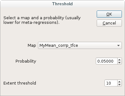
Results from the corresponding model can be thresholded to a given uncorrected or corrected statistical significance level, obtaining peak coordinates, clusters breakdowns and NIfTI images. This will also open MRIcron brain viewer to display the results (if MRIcron is enabled; see Settings section for help on setting up MRIcron integration).
Please find citation information for the atlas by Catani, Thiebaut de Schotten et al below.
One-tailed vs two-tailed
As in most neuroimaging software, this dialog conducts a pair of one-tailed tests. To avoid doubling the false positive rate, please halve the statistical significance level (e.g., to 0.025).
To threshold a map
Press the button [Threshold]
or:
Select [Threshold] in the Statistics menu, to open the following dialog:
Command-line and batch usage
threshold p_map_path,z_map_path,value,minimum number of voxels
Example:
threshold analysis_MyMean/corrp_tfce,analysis_MyMean/MyMean_z,0.05,10
Note: p-map and z-map paths should NOT include the extension .nii.gz
References
(effect-sizes and variances):
Radua J, Mataix-Cols D, Phillips ML, El-Hage W, Kronhaus DM, Cardoner N and
Surguladze S. A new meta-analytic method for neuroimaging studies that combines
reported peak coordinates and statistical parametric maps.
Eur Psychiatry 2012; 27:605–611.
![]() .
.
(white matter atlas 1):
Thiebaut de Schotten M, Dell'Acqua F, Forkel SJ, Simmons A, Vergani F,
Murphy DG and Catani M. A lateralized brain network for visuospatial
attention.
Nat Neurosci 2011; 14:1245-1246.
![]() .
.
(white matter atlas 2):
Thiebaut de Schotten M, Ffytche DH, Bizzi A, Dell'Acqua F, Allin M,
Walshe M, Murray R, Williams SC, Murphy DG and Catani M.
Atlasing location, asymmetry and inter-subject variability of white
matter tracts in the human brain with MR diffusion tractography.
Neuroimage 2011; 54:49-59.
![]() .
.
(white matter atlas 3):
Rojkova K, Volle E, Urbanski M, Humbert F, Dell'Acqua F and
Thiebaut de Schotten M. Atlasing the frontal lobe connections and their
variability due to age and education: a spherical deconvolution
tractography study.
Brain Struct Funct 2015; in Press.
![]() .
.